
 In clinical practice, cardiology is nowadays all about echocardiography. A physical examination, chest radiographs and an ECG will only get you so far. To make a definitive diagnosis, you need to be a master of echocardiography. And the best way to achieve that end is to do hands on training. Books will only get you half the way there; to get the whole way, you need one-on-one instruction with a gifted tutor using live animals.
The fastest way to make solid progress is to do basic course in echocardiography run by the CVE, and I recently enjoyed full weekend working with Niek Beijerink in Ross Pedrana’s practice in Dubbo. Ross kindly provided a venue, his own dogs and a range of canine patients with structural heart disease from his practice. John Marriot provided a range of up-to-date GE diagnostic ultrasound units, ultrasound cushions, ECG clips and abundant acoustic coupling gel. The whole weekend was devoted to teaching and learning, with equal emphasis on theory and practice. The first day was a “basic course” designed for people with limited experience with ultrasound. The second day was an “advanced course” for people who had some experience and expertise, but who wanted to take things “to the next level”.
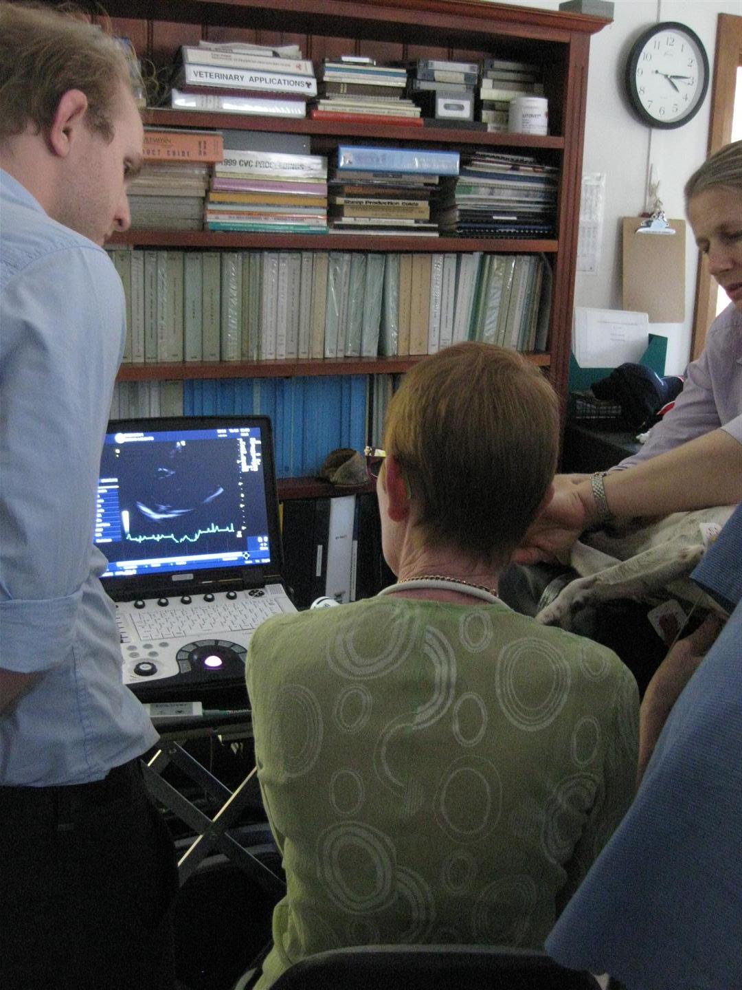
Niek is a skilled presenter and a very effective communicator. The first day started with the theory of diagnostic ultrasound, explaining the physics that underpins the practice of echocardiography. This is hard to make exciting, but Niek drew pertinent examples form real life to explain why you need a certain transducer frequency to do one application, but another to do something else. This was a good talk to start the day with, as it was hard work, yet completely essential. He then moved on to how to drive the machine, and how to obtain standard right parasternal long and short axis views, the bread and butter of echocardiography. This was first done in a series of PowerPoint presentations, and then again in real time using a normal canine “volunteer”. The delegates then got about 90 minutes of hand on scanning, working in groups if two at 6 different workstations, with Niek, his Resident and myself (as adjunct tutors) working with the 12 people attending the workshop. As each of the dogs had different forms of heart disease, and different thoracic wall conformation (and hence acoustic windows), the delegates moved from workstation to workstation during the course of the day. After lunch, Niek continued with further didactic presentations on M-mode echocardiography and the finer points of measurement, skills mandatory for quantitative recording of echocardiography data. This was reinforced by further demonstrations by Niek and another 90 minutes of hand-on scanning. Finally, to finish the day Niek went through a series of clinical cases with the delegates. This was a full on day, and I was pretty shattered at the end of it (it’s a long day starting at 9 am and finishing at 5.30), but the people attending were satisfied and happy, and felt they had achieved a lot. This included people that already had some considerable experience in diagnostic ultrasound; they felt they had “broken through” and now could do things faster and more reliably than before, and with better insight and understanding.
Six delegates stayed on for the second day of advanced training, which was even more “intimate” than the first day. Niek started with didactic lectures on principles of Doppler echocardiography, both spectral Doppler (PW and CW) and also colour-flow mapping. He then demonstrated these techniques on the patients. He then gave a detailed sessions on the views obtained from the left hemithorax (both caudal and cranial) and substernal windows, to show all the different ways the heart could be imaged, and how to optimise Doppler signals by tweaking the machines and optimising the views interrogated. There was over 3 hours of hands-on scanning time during the day, which ended up working through another challenging series of cases studies as a group. You can imagine with 6 delegates and 3 tutors, the people doing both day 1 and day 2 made amazing progress in terms of their scanning ability.
Niek is a very effective tutor. Perhaps because he is form the Netherlands, his use of English is very ordered and precise, and the slower delivery is actually very helpful to people listening to him speak (just ask any of his students from Sydney Uni!). He has a strong theoretical and practical knowledge, and is especially good at explaining difficult concepts, often by clever analogies. His passion for cardiology also comes through, which makes everything just a bit more worthwhile and fun. I love cardiology and have been doing echocardiography for over 20 years, but I learnt lots of new tricks and tips to add to my repertoire. On this particular weekend we had a wide range of vets attending – some form the city, some form the country, some young, some older, and two card-carrying diagnostic imagers (Annie Rose and Dayle Tyrrell). To a person, all delegates got a lot out of the weekend. Indeed, by the end of the sessions, there was quite camaraderie amongst the group. Niek was very generous with his time and his teaching material, providing all his power points to delegates go they could review them subsequently. John Marriot was happy as the machines performed at a high level providing terrific imagers (especially the high end units), and Ross Pedrana was happy to show off the excellent facilities at his Dubbo practice. It was a very satisfying weekend.
I cannot recommend this course more strongly. Cardiology is fun and it is lucrative if you do not need to refer all the patients for echocardiography. I would say after attending a course like this, and a few months of consolidation doing practice dogs and clinical cases, that most vets with some aptitude would be able to correctly diagnose 90 per cent of cardiac cases in their practices. This makes everyday practice more fun, and you will enjoy the kudos of being the go-to person for cardiology in your practice. If you can do four echoes a week, you can probably afford to pay the lease for a decent ultrasound machine, which of course you can use for many other purposes.
RICHARD MALIK
This article was provided by Radincon X-Ray.